| AnatOnMe - a virtual tool for physiotherapy |
| Written by Lucy Black | |||
| Sunday, 22 May 2011 | |||
|
Augmented Reality lets users look inside their body and see the location of anatomical detail. The idea is that by understanding what lies beneath people can better appreciate what they have to do to recover from injuries.
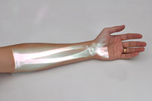
AnatOnMe is a device that projects images of the bones muscles and ligaments inside the body onto the patient's own skin and trials suggest it is helpful in motivating people recovering from injuries to do their exercises. This is a novel implementation of AR because instead of viewing the augmented world on a monitor or phone screen a projector is used to actually augment reality with a generated overlay! Microsoft Research demonstrated the prototype of AnatOnMe at this month's ACM CHI Conference on Human Factors in Computing Systems. It has been designed to help patients who are undergoing physiotherapy understand their injuries and thus encourage them to stick to the exercise regime and reduce the degreee of non-compliance that explains the high failure rate of this type of therapy. According to Amy Karlson, of Microsoft Research's Computational User Experiences Group in Redmond, Washington: "People are notoriously bad at sticking to their physical therapy regimens. Between 30 and 50 percent of patients with chronic conditions fail to comply with their recommended therapies and a s a result, conditions can take longer to heal or can get even worse". However, the more information that patients have about their injuries, the more likely they are to comply with physical therapy regimens. Designed to provide this information AnatOnMe, projects an image of the underlying bone structure, muscle tissue, tendons, or nerves onto the skin, giving patients a better understanding of the injury, and of what they need to do to help the healing process.
"The technology is somewhat low-tech," commented Karlson explainaing how the prototype device comes in two parts. The first contains a handheld, or pico, projector, an ordinary digital camera, and an infrared camera. The second contains a laser pointer and the control buttons. Instead of using a complicated autocorrection system to map the image of the internal injury precisely onto the patient's exterior, the therapist simply points the projector and lines it up by eye. Also with the prototype, the images displayed are not actually taken from scans of the patients but come from stock graphical images used to show one of six different types of injury.
Despite these shortcomings, controlled experiments of the device carried out by two physical therapists suggest that the device encourages patients to stick to their therapies.Hopefully this will be sufficient to improve on the design of the prototype and produce a tool that uses scans taken from an individual patient. Writing on Fast Company, David Zax writes: "Imagine if a truly smart version of this technology used an augmented reality app to recognize the shape of a patient's leg and display an animation of the underlying mechanics during a workout. Imagine if that app were integrated with the data the doctor acquires on each visit, so that it would be updated with your progress as you continue your regimen.If the low-tech ideas here got real high-tech treatment, non-compliance might just go the way of polio."
|
|||
| Last Updated ( Sunday, 22 May 2011 ) |


